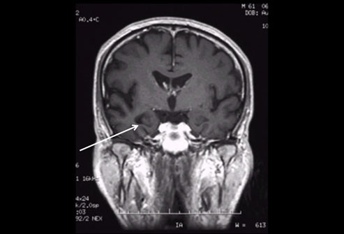
DEMENTIA
Q1.All are neuroimaging feature suggestive of dementia exept
a.Alzheimer’s disease---hippocampal atrophy and posterior dominant cortical atrophy
b.Fronto-temporal lobe degenaration(FTD)—frontal,insular and or temporal atrophy,spares posterior parietal lobule
c.Dementia with Lewy body(DLB)—posteror parietal atrophy,hippocampi larger than AD,greater involvemet of amygdale than hippocampi
d.Cruetzfeld -Jacob diseases(CJD)—cortical ribboning , basal ganglia or thalamus hypointensity on diffusion or FLAIR MRI
ANS---d===Cruetzfeld -Jacob diseases(CJD)—cortical ribboning, basal ganglia or thalamus hyperintensity on diffusion or FLAIR MRI
Alzheimer’s disease
- the most common cause of primary degenerative dementia,
- incidence increases with age, rising sharply over 70 years.
- It is a generalized disorder, although in its early stages it may affect the medial part of the temporal lobe, especially the hippocampus, more than elsewhere.
Structural imaging
- This is usually normal, although occasionally accentuated atrophy medially in the temporal lobe is indicated by widening of the perihippocampal CSF spaces, which should be both bilateral and symmetrical
- In established disease, characteristic symmetrical posterior temporal and parietal perfusion defects on regional cerebral blood flow (rCBF) SPECT have a predictive value for the diagnosis of AD of over 80% and the severity of rCBF reduction correlates with the degree of cognitive decline
- [18F]deoxy-d-glucose (FDG)-positron emission tomography (PET) studies show reduced glucose uptake that is not explained by atrophy
- Receptor imaging using radioligands for central benzodiazepine receptors has shown a similar distribution of deficits in AD
Frontotemporal dementia
|
- These comprise less than 10% of the primary degenerative dementias. They include Pick's disease.
- MRI and even CT show atrophy in the anterior and medial parts of the temporal lobe, which usually is markedly asymmetric (right or left) and diminishes posteriorly. Asymmetric frontal lobe atrophy may also be present
Functional imaging
| |
- Reduced frontal perfusion is not specific to frontotemporal dementia and can occur in a variety of other conditions, such as schizophrenia, depression, human immunodeficiency virus (HIV) encephalopathy, Creutzfeldt–Jacob disease (CJD) and in some cases of AD
- Lewy body dementia is now recognized as the second most common degenerative dementia after AD and it accounts for approximately 20% of all dementia.
- Neither structural imaging nor rCBF SPECT can reliably distinguish between Lewy body dementia and AD on subjective assessment, as posterior temporoparietal defects occur in both. However, reduced frontal perfusion with HMPOA SPECT and reduced uptake in the cerebellum and visual cortex with FDG-PET is seen in Lewy body dementia compared with AD.
- This is the clinical diagnosis in about 20% of all dementias. Evidence of ischaemic damage on CT or MRI is mandatory for diagnosis,
- These mainly comprise CJD (sporadic, iatrogenic, familial) and very recently in Europe (especially in the UK) new variant CJD (nvCJD). Rapidly progressive dementia, often with myoclonus, is the usual clinical picture, often preceded by behavioural disturbances, especially in nvCJD.
- Structural imaging usually appears normal in early stages, but rapidly progressive atrophy soon develops.
- Symmetrical increases in signal in the putamen and caudate nuclei may be shown by MRI in about 10% of sporadic CJD, and in the posterior part of the thalami in over 50% of nvCJD;
No comments:
Post a Comment