Friday 26 June 2015

Molecular breast imaging (MBI)


New research from Mayo Clinic demonstrates that using molecular breast imaging (MBI) as an adjunct to mammography results in an almost four-fold increase in invasive cancer detection in women with dense breast tissue. These findings, along with results of earlier studies, have spurred the Rochester, Minn., clinic to make supplemental imaging with MBI its standard of care for women with dense breasts.
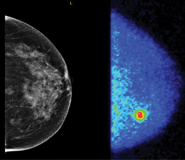
MBI utilizes a small gamma camera to acquire images of the breast after injection of the radiotracer sestamibi.
"Breast tumors very avidly take up sestamibi," said Dr. O'Connor, who collaborated on the research with radiologists, surgeons and physicians at the Breast Diagnostic Clinic at Mayo Clinic in Minnesota. "With this technique, we're not looking at the architecture of the lesion, but rather its metabolic activity."
Breast imaging expert Wendie Berg, M.D., Ph.D., professor of radiology at Magee-Womens Hospital of the University of Pittsburgh School of Medicine, said the findings show a potential role for MBI in screening the approximately 40 percent of women over age 40 who have dense breasts.

Monday 22 June 2015

MCQ ---Neuro-module

An adult female patients complain of isolated pain in right eye  with features of ptosis, diplopia, pupillary dilation, and strabismus.The symptom persist with no evidence of improvement .The clinician suspected of intracranial aneurysm and so the patient was advised for CT ANGIORAPHY.The most likely site of aneurysm in such case is
a. The junction of the anterior cerebral and anterior communicating arteries
b. the ICA at the origin of the posterior communicating artery
c.the bifurcation of the MCA
d.the posterior cerebral artery

e. the bifurcation of the ICA

Thursday 18 June 2015

MCQ ---CAVERNOUS ANGIOMA


112.MR features of cavernous angioma is /are

a. focal central heterogeneity containing areas corresponding to subacute-chronic hemorrhage (methemoglobin)
b. circumferential complete rings of markedly hyperintense iron-storage forms around low -intensity central areas
c. no mass effect or edema
d. no demonstrable feeding arteries or draining veins associated with developmental venous anomalies

e. contrast enhancement

Saturday 13 June 2015

MCQ --NEURORADIOLOGY---Moyamoya disease


101.All are true regarding Moyamoya disease except
a. progressive symmetric occlusion involving the bifurcations of the internal carotid arteries (ICAs) and the proximal anterior and middle cerebral arteries
b. development of an extensive network of enlarged basal, transcortical, and transdural collateral vessels.
c. presence  of the expected flow void within the cavernous and supraclinoid portions of the ICAs
d. The angiographic appearance of the innumerable tiny collateral vessels, termed “puff of smoke” or “moyamoya” in Japanese.

e. Moyamoya disease has a bimodal age presentation, with the first peak occurring in the first decade of life, associated with cerebral infarction


105.What is the size of circle of Willis aneurysms  that can be detected in most of cases  by intracranial MRA  by  state-of-the-art acquisition and postprocessing methodology
a.>2mm
b. >3mm
c. >4mm
d. >5mm

e. >6mm

ANS---
101---c
Moyamoya disease has a bimodal age presentation, with the first peak occurring in the first decade of life, associated with cerebral infarction as progressive carotid occlusion develops. Adult patients most often present in the fourth decade with intracranial hemorrhage arising from the rupture of the delicate network of collateral vessels
Absence of the expected flow void within the cavernous and supraclinoid portions of the ICAs is a consequence of narrowing and ultimately occlusion of these vessels

There is increased incidence of moyamoya changes in patients with Down syndrome.
105---b

Sunday 7 June 2015

FRCR MCQ ---NEURO MUDULE (INFARCTION)


What is approximate age of infarction in A and D image?


ANS—

A.-- ADC values seen to be low within hours after the stroke and continue to decline for the next few days. They remain reduced through the first 4 to 5 days after stroke and then undergo pseudonormalization between 4 and 10 days . After this the ADC subsequently has been found to rise in the lesion (i.e., the ADC map shows hyperintensity) beyond 10 days  So,approximate age of infarction in A is less than 4—5 days while that of D is about more than ten days.

Thursday 4 June 2015

DIAGNOSIS ?


WHAT IS FINDING IN ABOVE MRI IMAGE? WHAT IS DIAGNOSIS?

ANS---

The lesion in right parietal lobe  shows central hyperintensity with peripheral circumferential rims of hypointensity  on both T1W and T2W images.The hypointensity becomes more prominent on GRE s/ of hemosiderin/ferritin.There is no edema.All these features are nearly specific for cavernous angioma.

Monday 1 June 2015

FRCR MCQ ----NEURO MODULE


1.From above MRI image ,answer following questions
A.Name MRI the sequence of a,b,c,d,e
B.What is the location of lesion?
C.What is finding in a,b.c,d.e?
D. What is diagnosis and why ?


ANS---
A.—
a.---T1W image
 b.—T2W image
 c.-- gradient recalled echo image
 d.-- the diffusion-weighted image
 e.--- the calculated ADC map
B.The location of lesion is left basal ganglia
C.----
a.---hypointense to isointense to brain on T-weighted images
b.---hyperintense to brain on T2-weighted images with a peripheral rim of marked hypointensity
c.---GRE shows hypointensity of lesion
d.---the lesion is hyperintense on diffusion weighted image
d.---reduced ADC map
Hematomas containing intracellular oxyhemoglobin (OxyHbIntra), intracellular deoxyhemoglobin (DeoxyHbIntra), and intracellular methemoglobin (MetHbIntra) all have restricted diffusion (reduced ADC), whereas hematomas with extracellular (“free”) methemoglobin (MetHbFree) have elevated ADC
D.
Hyperacute hematoma of left basal ganglia
Hyperacute hematomas appear slightly hypointense or isointense to brain on T-weighted images and slightly hyperintense to brain on T2-weighted images. A thin, irregular rim of marked hypointensity at the periphery of the lesion on T2-weighted images  is paramount to recognize it. This has been attributed to very rapid deoxygenation of blood within the hematoma at the blood–tissue interface. The hypointensity may be more evident in 3-T than lower-field systems and more obvious on GRE images regardless of field strength; regardless of scanner field strength.So, T2*-weighted GRE should be part of all routine stroke MR protocols.
Restricted diffusion and reduced ADC map is due to presence of oxyhemoglobin