| Head |
|
|
|
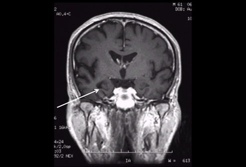
| Cerebrovascular accident |
Cerebral perfusion SPECT |
99mTc HMPAO |
Uptake proportional to blood flow |
| Hydrocephalus |
![]() |
|
|
|
| CSF rhinorrhoea |
Cerebrospinal fluid (CSF) study |
111In DTPA (intrathecal) |
Marker of CSF flow. |
| Encephalitis |
Blood–brain barrier (BBB) study |
99mTc HMPAO |
Passage across disrupted BBB |
| Dementia |
Cerebral perfusion SPECT |
99mTc HMPAO |
Uptake proportional to blood flow |
|
|
Cerebral metabolism PET |
18F fluorodeoxyglucose |
Marker of glucose metabolism |
| Epilepsy (presurgical localization) |
Ictal SPECT |
99mTc HMPAO |
Uptake proportional to blood flow |
|
|
Interictal PET |
18F fluorodeoxyglucose |
Marker of glucose metabolism |
| Neck |
|
|
|
| Thyrotoxicosis |
|
|
123I sodium iodide |
Active uptake (123I and 99mTc) followed by
organification (123I) |
| Thyroid nodule |
![]() |
Thyroid scintigraphy |
99m Tc pertechnetate |
|
| Ectopic thyroid |
|
|
|
| Hyperparathyroidism (presurgical localization) |
Parathyroid scintigraphy |
99mTc MIBI |
Differential expression of p-glycoprotein between parathyroid
adenoma and thyroid |
| Dry mouth (connective tissue disease) |
Salivary gland study |
99mTc pertechnetate |
Secretion in saliva |
| Musculoskeletal system |
|
|
|
| Tumour |
![]() |
|
|
|
| Fracture |
|
|
|
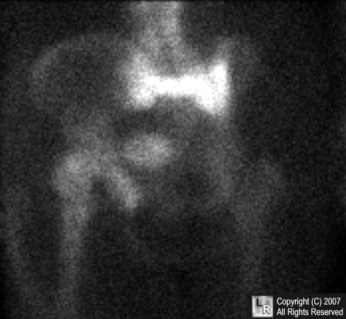
| Avascular necrosis |
Bone scintigraphy |
99mTc polyphosphate compounds |
Osteoblastic response (+ vascularity on early phases) |
| Arthropathy |
|
|
|
| Metabolic bone disease |
|
|
|
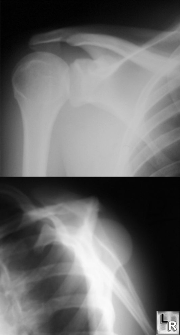
| Painful prosthesis |
![]() |
|
|
|
| Osteomyelitis |
|
99mTc polyphosphate |
Osteoblastic activity |
|
|
Bone scintigraphy + white cell or gallium scintigraphy |
99mTc- or 111In-leucocytes |
Leucocyte migration |
|
|
|
67Ga gallium citrate |
Binds to transferrin and leaks into extravascular space |
| Lymphoedema |
Lymphoscintigraphy |
99mTc nanocolloid |
Lymphatic uptake and trapping |
| Cardiovascular system |
|
|
|
| Chest pain |
Myocardial perfusion scan |
201Tl (thallous chloride) |
K+ analogue indicating perfusion (ischaemic heart disease)
(delayed uptake reflects viability) |
|
|
|
99mTc isonitriles |
Cationic complexes taken up by myocytes in proportion to blood
flow |
|
|
|
99mTc teboroxime |
Lipophilic compound which accumulates by diffusion |
|
|
|
99mTc phosphines |
Uptake proportional to blood flow |
| Cardiac failure |
Cardiac ventriculography (gated study) |
99mTc red blood cells |
Blood pool label |
|
|
Myocardial viability study |
18F fluorodeoxyglucose |
Demonstrates shift from metabolism of fatty acids to
glucose |
| Pulmonary embolism |
Ventilation/perfusion (V/Q) scan |
123I fatty acids |
|
|
|
|
Perfusion: 99mTc albumin |
Pulmonary arteriole blockade |
|
|
|
Macroaggregates |
|
|
|
|
Ventilation: 99mTc aerosols |
Distributes in lungs in proportion to gas regional
ventilation |
|
|
|
133Xe gas, 81mKr gas |
|
| Congenital heart disease |
Quantitative shunt study |
99mTc red blood cells |
Blood pool label |
| Pulmonary system |
|
|
|
| Solitary pulmonary nodule |
Tumour imaging |
18F fluorodeoxyglucose |
Marker of glucose metabolism |
| Occult lung disease (alveolitis) |
Alveolar permeability study |
99mTc DPTA aerosol |
Passage across alveolar membrane into blood |
| Gastrointestinal system |
|
|
|
| Difficulty in swallowing |
Oesophageal transit and reflux |
99mTc sulphur colloid |
Transit of labelled material |
| Gastrointestinal haemorrhage |
GI bleed study |
99mTc sulphur colloid |
Blood pool label extravasating into bowel |
|
|
|
99mTc labelled red cells |
|
| Ectopic gastric mucosa |
Meckel's diverticulum scintigraphy |
99mTc pertechnetate |
Active uptake by ectopic gastric mucosa |
| Diarrhoea (inflammatory bowel disease) |
White cell scintigraphy |
99mTc leucocytes |
Leucocyte migration |
| Vomiting (gastroparesis) |
![]() |
|
99mTc sulphur colloid in egg (solid phase) |
|
|
|
Gastric emptying study |
|
Compartmental localization of labelled material |
| Dumping |
|
|
111In DTPA in orange juice (liquid phase) |
|
| Focal liver lesion (haemangioma) |
Red blood cell study |
99mTc labelled red blood cells |
Red cell pooling |
| Cholecystitis |
![]() |
|
|
|
| Biliary dyskinesia |
|
|
|
|
Hepatobiliary study |
99mTc iminodiacetic acid derivatives |
Uptake by hepatocytes and excretion into bile |
| Biliary atresia |
|
|
|
| Bile leak (post-op) |
|
|
|
| Abdominal sepsis |
![]() |
|
99mTc or 111In leucocytes |
Leucocyte migration |
|
White cell or gallium scintigraphy |
|
|
| Pyrexia of unknown origin |
|
67Ga gallium citrate |
Binds to transferrin and leaks into extravascular space |
| Ectopic splenic tissue |
Splenic scintigraphy |
Heat damaged 99mTc labelled red blood cells |
Splenic trapping of damaged cells |
| Urological, adrenal and genitourinary
systems |
| Hypertension (renovascular disease) |
Captopril renography |
99mTc MAG3 |
Captopril-induced change in renal transit time and/or
function |
| Renal tract obstruction |
Diuresis renography |
99mTc DTPA |
Glomerular filtration |
|
|
|
99mTc MAG3 |
Proximal tubular secretion |
| Renal scarring |
Static renal scintigraphy |
99mTc DMSA |
Glomerular filtration and proximal tubular uptake |
| Vesicoureteric reflux |
Indirect micturating cystogram |
99mTc MAG3 |
Compartmental localization |
| Adrenal medullary tumour |
Adrenal study |
123I MIBG |
Uptake by noradrenaline transporter |
| Adrenal cortical tumour |
Adrenal study |
123I iodocholesterol |
Incorporation into hormone metabolism |
| Cancer |
| Space occupying lesion in brain (SOL) |
Tumour imaging |
201Tl (thallous chloride) |
K+ analogue indicating perfusion |
|
|
|
18F fluorodeoxyglucose |
Marker of glucose metabolism |
| Thyroid cancer |
Whole body iodine scintigram |
131I sodium iodide |
Uptake by Na/I transporter |
| Skeletal metastases |
Bone scintigraphy |
99mTc polyphosphate |
Osteoblastic response |
| Soft tissue mass (sarcoma) |
Tumour imaging |
201Tl (thallous chloride) |
K+ analogue indicating perfusion |
|
|
|
18F fluorodeoxyglucose |
Marker of glucose metabolism |
| Tumour staging |
![]() |
|
|
|
| Tumour recurrence |
Tumour imaging |
18F fluorodeoxyglucose |
Tumour glucose metabolism |
| Tumour response assessment |
|
|
|
| Insulinoma |
![]() |
Somatostatin receptor study |
111In pentetreotide (Octreotide®) |
Binds to somatostatin receptors |
| Carcinoid tumour |
|
|
|
| Neuroblastoma |
MIBG scintigram |
123I MIBG |
Uptake by noradrenaline transporter |
| Tumour hypoxia |
Hypoxia imaging |
18F fluoromisonidazole |
Trapped in hypoxic cells |
| Sentinel node detection |
Lymphoscintigraphy |
99mTc nanocolloid |
Lymphatic uptake and trapping |