Friday 26 September 2014

ADHD ------MR IMAGING

Multimodal MR Imaging of Brain Iron in Attention Deficit Hyperactivity Disorder: A Noninvasive Biomarker That Responds to Psychostimulant Treatment?

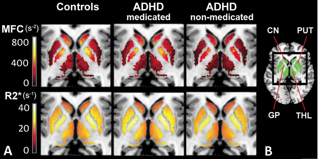
Lower magnetic field correlation (MFC) indexes of striatal and thalamic brain iron in medication-naïve attention deficit hyperactivity disorder (ADHD) patients and lack of differences in psychostimulant-medicated patients suggest that MFC indexes of brain iron may represent a noninvasive diagnostic biomarker that responds to psychostimulant treatment
Vitria Adisetiyo, Ph.D., of the Medical University of South Carolina, Charleston, and colleagues prospectively examined 22 patients with ADHD (12 medication-naïve patients and 10 with a history of psychostimulant treatment) and 27 control subjects (age range, 8 to 18 years). Brain iron was indexed noninvasively using MR imaging relaxation rates (R2, R2*, R2') and MFC in the globus pallidus, putamen, caudate nucleus and thalamus. Serum iron measures were also collected. Subgroup differences were analyzed with data-appropriate omnibus tests followed by post-hoc pairwise comparisons; false discovery rate correction was conducted to control for multiple comparisons.
Medication-naïve ADHD patients had significantly lower striatal and thalamic MFC indexes of brain iron than control subjects (putamen, P = .012; caudate nucleus, P = .008; thalamus, P = .012) and psychostimulant-medicated ADHD patients (putamen, P = .006; caudate nucleus, P = .010; thalamus, P = .021). Conversely, the MFC indexes in medicated patients were comparable to those in control subjects. No significant differences were detected with R2, R2*, R2' or serum measures.
“Our results implicate reduced striatal and thalamic brain iron levels in ADHD pathophysiology before medication and suggest that reduced brain iron levels may normalize with psychostimulant treatment,” the authors write.


Media Coverage of RSNA

Sunday 21 September 2014

World Alzheimer’s Day: September 21

World Alzheimer’s Day: September 21

World Alzheimer's Day, September 21st of each year, is a day on which Alzheimer's organizations around the world concentrate their efforts on raising awareness about Alzheimer's and dementia. Alzheimer's disease is the most common form of dementia, a group of disorders that impairs mental functioning.

Thursday 18 September 2014

Reversed halo sign /Atoll sign

Reversed halo sign /Atoll sign

The reversed halo sign (also know as the atoll sign, reverse halo sign, and the fairy ring sign) is defined by central ground-glass opacity with a surrounding halo or crescent of consolidation .

This should not be confused with the halo sign which refers to central consolidation with a surrounding halo or rim of ground-glass opacity.

Furthermore, the reversed halo sign should be differentiated from cavitation (development of focal regions of internal gas) or pseudocavitation (likely internal bronchiolectasis described in subtypes of lung adenocarcinoma).

The reversed halo sign was first described and is most commonly associated with COP but is not specific to this disease. It has been reported in the setting of bacterial pneumonia, mucormycosis, paracoccidioidomycosis, tuberculosis, sarcoidosis, radiofrequency ablation, lymphomatoid granulomatosis, Wegener granulomatosis, tumor, and pulmonary infarcts.


.


Figure 1
Figure 1
Image Tools


Friday 12 September 2014

SPARROW TAIL APPEARANCE OF SUSTANTIA NIGRA IN PARKINSONISM


  • Recent advances in MR imaging point the way to a reliable marker for Parkinson disease, which is usually diagnosed almost solely through medical history and clinical findings like muscle stiffness and tremors.
  • Three MR imaging-based studies have discovered distinctive changes in the substantia nigra (SN), a crescent-shaped mass of cells in the midbrain that normally produces the neurotransmitter dopamine. 
  • Parkinson disease patients lose dopamine-producing cells in the SN, leading to problems with motor control among other symptoms. All three studies showed consistent differences in the appearance of the SN in normal patients compared with patients diagnosed with Parkinson disease.
  • The initial study was conducted by neuroradiologists, neurologists and physicists of the University of Nottingham in the U.K., and published in the July 2013 edition of Neurology. Led by resesarchers Anna Blazejewska, Ph.D., and Stefan Schwarz, M.D., the team investigated 7-T MR imaging changes to nigrosome-1 of the substantia nigra revealing an oval shaped structure found in healthy patients but absent in patients with Parkinson disease.
  • In a follow-up study by the same research team led by Dr. Schwarz, previous findings were translated to a 3-T MR imaging platform revealing a specific “swallow tail” shape indicating the presence of nigrosome-1—part of the SN that has a dense concentration of dopamine-producing cells. Researchers determined that the split-tail shape was clearly visible in normal patients but not present in the patients with Parkinson disease.


RSNA NEWS

Wednesday 10 September 2014

MCQ ----FOR PG EXAM

55.All are true regarding mature teratoma in mediastinum except

a.m/c germ cell tumour

b.mostly solid

c.may have fat

d.may have calcification


e.located in anterior mediastinum

57.All are true regarding mediastinal lymphnode calcification except

a. common following tuberculosis and fungal infection

b.  common in metastatic neoplasm

c. may be seen in lymph node metastases osteosarcoma, chondrosarcoma

d.  foamy appearance seen with Pneumocystis jiroveci  infection  in AIDS patients

e. eggshell calcification in sarcoidosis 

Tuesday 9 September 2014

RADIOLOGY MCQ FOR PG

1.The most sensitive investigation to diagnose renal TB in early stage is IVP
TRUE /FALSE

2.
COBRA HEADED appearance on IVP is seen in vesicocoele
True /false

3.Investigation of choice for urolithiasis is CECT
true /false

4.Dromedary hump is related to diaphragm 
TRUE/FALSE

5.COBRA HEADED appearance on IVP is seen in vesicocoele
True /false


WRITE ANS IN COMMENTS-------

Wednesday 3 September 2014

MCQS ---FRCR AND PG---- CHEST MODULE

30. Sign almost invariably present with significant tension pneumothorax is

a.ipsilateral mediatinal shift

b.ipsilateral diaphragmatic depression

c. contralateral  diaphragmatic depression

d. Double diaphragm sign


e. ipsilateral mediatinal shift

33.All are true regarding fibrothorax /diffuse pleural thickening

a. a smooth uninterrupted pleural density that extends over at least one-quarter of the chest wall

b. extends more than 8 cm in the craniocaudal direction, 5 cm laterally and  a thickness of more than 3 mm on CT scan

c. Extensive calcification favours previous tuberculosis or empyema

d. Asbestos exposure-related fibrothorax is usually bilateral and heavily calcified

e. mesothelioma, metastatic disease (particularly adenocarcinoma), lymphoma and leukaemia may cause fibrothorax

ANS BELOW----

FRCR PART 1 MCQs  Radiophysics (e-book)

FRCR PART 1 MCQs Radiophysics (e-book)

Conventional Radiography Computed Radiography Digital Radiography Gamma imaging MRI USG

by Dr.Nagendra Kumar Sinha MD (Radio-diagnosis) (write a review)
Type: e-book
Genre: Diet & Health, Medicine & Science
Language: English
Price: Rs.1,177.00
Available Formats: PDF Immediate Download on Full Payment
Preview the book   
- See more at: http://pothi.com/pothi/book/ebook-dr-nagendra-kumar-sinha-md-radio-diagnosis-frcr-part-1-mcqs-radiophysics#sthash.CLtvh6HI.dpuf


ANS
30.Moderate or gross mediastinal shift  should be taken as 

indicating tension, particularly if the ipsilateral hemidiaphragm is

 depressed. Depreesed ipsilateral hemidiaphragm  is the more

 reliable and is almost invariably present with significant tension 

pneumothorax. 

33.Asbestos exposure-related fibrothorax is less common than

 pleural plaques and is usually the sequel of a benign exudative

 effusion. Asbestos exposure-related fibrothorax is usually 

bilateral and rarely calcified.