Tuesday 10 December 2013

Brockenbrough-Braunwald sign


Brockenbrough-Braunwald sign

Q1.Brockenbrough-Braunwald sign noted during cardiac catheterization is seen in
a.aortic  stenosis
b.hypertrophic obstructive cardiomyopathy
c.mitral stenosis

d.pericarditis

ANS.-----b====Brockenbrough-Braunwald sign noted during cardiac catheterization is seen in hypertrophic obstructive cardiomyopathy


  • Upon cardiac catheterizationcatheters can be placed in the left ventricle and the ascending aorta, to measure the pressure difference between these structures.
  •  In normal individuals, during ventricular systole, the pressure in the ascending aorta and the left ventricle will equalize, and the aortic valve is open. 
  • In individuals with aortic stenosis or with HCM with an outflow tract gradient, there will be a pressure gradient (difference) between the left ventricle and the aorta, with the left ventricular pressure higher than the aortic pressure. This gradient represents the degree of obstruction that has to be overcome in order to eject blood from the left ventricle.
  • The Brockenbrough–Braunwald–Morrow sign is observed in individuals with HCM with outflow tract gradient. This sign can be used to differentiate HCM from aortic stenosis.
  •  In individuals with aortic stenosis, after a premature ventricular contraction (PVC), the following ventricular contraction will be more forceful, and the pressure generated in the left ventricle will be higher. Because of the fixed obstruction that the stenotic aortic valve represents, the post-PVC ascending aortic pressure will increase as well. 
  • In individuals with HCM, however, the degree of obstruction will increase more than the force of contraction will increase in the post-PVC beat. The result of this is that the left ventricular pressure increases and the ascending aortic pressuredecreases, with an increase in the LVOT gradient.
  • While the Brockenbrough–Braunwald–Morrow sign is most dramatically demonstrated using simultaneous intra-cardiac and intra-aortic catheters, it can be seen on routine physical examination as a decrease in the pulse pressure in the post-PVC beat in individuals with HCM.


Monday 9 December 2013

Ollier’s disease

Ollier’s disease/Maffuci’syndrome/Beckwith- Widerman’s syndrome /Klippel-trenaunay-Weber  syndrome


Q1.Enchondrma and heamangioma is found in
a.Ollier’s disease
b.Maffuci’syndrome
c.Beckwith- Widerman’s syndrome

d.Klippel-trenaunay –Weber  syndrome


ANS. b===MAFFUSI SYNDROME------ Maffucci syndrome is a congenital non hereditary mesodermal dysplasia  characterised by multiple enchondromas with soft  tissue cavernous haemangiomas


  • Ollier disease ( enchondromatosis) is a non-hereditary, sporadic, skeletal disorder characterised by multiple enchondromas that are principally located in the metaphyseal regions.


  • Beckwith–Wiedemann syndrome  is an overgrowth disorder usually (but not always) present at birth characterized by an increased risk of childhood cancer and certain congenital features.
  •  Five common features used to define the syndrome are: macroglossia, macrosomia (birth weight and length greater than the 90th percentile), midline abdominal wall defects (omphalocele/exomphalos, umbilical hernia, diastasis recti), ear creases or ear pits, and neonatalhypoglycemia (low blood sugar after birth).


Sunday 8 December 2013

FRCR MCQ ---MRI (Inversion recovery)


MRI (Inversion recovery)


Q1. The MRI technique used to null signal from fluids is
a.FLAIR
b.STIR
c.GRE
d.HASTE

Q2.The MRI technique used to null signal from fat is
a.FLAIR
b.STIR
c.GRE
d.RARE

ANS.Q1===a----The MRI technique used to null signal from fluids                              is FLAIR(fluid attenuated inversion recovery )
        Q2===b---- The MRI technique used to null signal from fat is                               STIR(short-tau inversion recovery)


Inversion recovery

  • In this sequence type, the magnetization is ‘prepared’ before the initial 90-degree excitation pulse by the addition of an 180-degree ‘inversion’ pulse. 
  • The signal obtained will be influenced by the relative degree of recovery experienced by the spins along the z-axis. 
  • The inversion time (TI) is the time allotted for this recovery process to evolve between the 180-degree inversion and 90-degree excitation pulses. The inversion pulse introduces heavy T1 weighting into this sequence.

Specific forms of the technique -


www.mriworld.org


  • A== fluid attenuated inversion recovery (FLAIR) where the TI is chosen so that the magnetization from fluids is nulled (≈2200 ms) in the detection (x–y) plane and TE is long (80–100 ms), introducing a lot of T2 as well as T1 weighting. Thecombination  of fluid nulling with T2 weighting is particularly useful in assessing lesions with prolonged T2 adjacent to fluid structures (e.g. periventricular demyelination).


  •  B====In short-tau inversion recovery (STIR), T1 is kept short, and is typically used to null signal from fat. An example of its use in this context is in the delineation of breast lesions and orbital pathology. 


Friday 6 December 2013

RADIOLOGY MCQ----Drooping lily sign


Drooping lily sign

Q1.Drooping lily sign on IVU is seen in
a.Duplex kidney
b.Hydatid cyst of kidney
c.APKD
d.Multicystic kidney

ANS --1===Drooping lily sign on IVU is seen in Duplex kidney


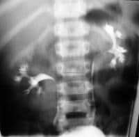
  • Drooping lily sign is  a urographic sign  of duplicated  renal collecting system
  • It refers to the inferolateral displacement of the opacified lower pole moiety due to an obstructed (and relatively unopacified) upper pole moiety. The similarity to a lily is further strengthened by the small number of calyces the lower pole moiety has 
  • In duplicated collecting system it is classically the upper pole ureter that is obstructed due to a ureterocoele and
  •  the lower pole ureter that refluxes: as described by the Weigert-Meyer law


radiopaedia.org


emedicine.medscape.com

Thursday 5 December 2013

RADIOLOGY MCQ---Lemon sign and Banana sign


Lemon sign and Banana sign

Q1.Banana sign refers to 
a.flattening of cerebellar hemisphere
b.scalloping of frontal bones
c.flattening of cerebrum
d.none

Q2.Lemon sign and banana sign are found in 
a.open spina bifida
b.Arnold-Chiari malformation
c.both
d.none

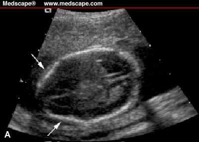
ANS.1===a==Banana sign refers to flattening of cerebellar                                    hemisphere
         2===c==Lemon sign and banana sign are found in .open                                 spina bifida and Arnold-Chiari malformation


The banana sign refers to the shape of the cerebellum

 owing to caudal displacement; the lemon sign refers to the

 lemon-shaped head resulting from scalloping of the frontal

 bones.


Wednesday 4 December 2013

RADIOLOGY MCQ---Prune- belly syndrome


Prune- belly syndrome

Q1.Prune- belly syndrome is characterized by all except
a.bilateral hydronephrosis and hydroureter
b.distended bladder
c.undescended testes
d.normal anterior abdominal wall

ANS--d===normal anterior abdominal wall

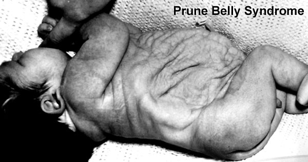
  • ThePrune- belly syndrome   (PBS) is generally readily apparent clinically and consists of absent abdominal wall muscles, undescended testes, and renal dysplasia associated with gross dilatation of the collecting systems.
  • All affected children have a universal mesentery with a mobile bowel.
  • Plain abdominal radiographs show a protuberant abdomen resulting from the lack of abdominal musculature. In the most severely affected patients, there may be urethral obstruction with severe renal dysplasia, while less severely affected individuals demonstrate an array of urinary tract abnormalities.
  • The characteristic features are small kidneys with abnormal minimally dilated calyces and upper ureters, while the lower ureters are tortuous and show disproportionate dilatation.
  •  The bladder is thin walled, of large capacity without trabeculation, and has a wide neck.
  •  A patent urachus or a urachal diverticulum may be present.
  • The posterior urethra is dilated proximally with a typical conical narrowing and a poor stream in the distal urethra .
  •  Occasionally, generalized or focal anterior urethral dilatation is present. Prognosis depends on the degree of renal dysplasia.


php.med.unsw.edu.au


www.pedsradiology.com


REF

Adam: Grainger & Allison's Diagnostic Radiology, 5th ed.

Tuesday 3 December 2013

RADIOLOCY MCQ--Sunburst sign / T sign / Lambda sign

Sunburst sign / T sign / Lambda sign


Q1.T- sign at two amnion junctions suggests of
a.dichorionic-diamniotic  twin
b.monochorionic-diamniotic  twin
c.monochorionic-monomniotic  twin
d.all
Q2.Lambda/Twin peak sign in USG of pregnancy is noted in
a.dichorionic-diamniotic  twin
b.monochorionic-diamniotic  twin
c.monochorionic-monomniotic  twin
d.all
Q3.Radial arrangement of medial sulci above the third ventricle(sunburst sign) on usg of cranium is seen in
a.corpus callosum agenesis
b.spinan bifida
c.Arnold-chiari malformation
d.Dandy-Walker malformation

ANS   Q1===b ------.monochorionic-diamniotic  twin
           Q2===a--------dichorionic-diamniotic  twin
           Q3===a-----corpus callosum agenesis


Monday 2 December 2013

RADILOGY MCQ--Acromegalyand Hyperthyroidism

RADIOLOGICAL FEATURES OF ACROMEGALY,CUSHING SYNDROME,HYPERTHYROIDISM AND HYPOTHYROIDISM


Q1.Tufting of the terminal phalanges (arrow head appearance)  and increased heel pad thicknes is seen in
a.alkaptonuria
b.hyperthyroisism
c.acromegaly

d.cushing syndrome


Q2.Small or absent frontal sinuses are noted in all except
a.congenital absence
b.hyperthyroidism
c.Down’s syndrome
d.kartagener’s syndrome

ANS---1==c ===acromegaly
             2==b===hyperthyroidism


  RADIOLOGICAL FEATURES OF ACROMEGALY


Skull Vault thickened
Paranasal and mastoid air cells enlarged
Pituitary fossa enlarged
Floor of the fossa asymmetrical or ballooned
Mandible Prognathism with increased angle
Spine Kyphosis
Enlarged vertebral bodies
Posterior scalloping of vertebral bodies
Chest Increased antero-posterior diameter
Ribs increased in calibre and length
Hands General enlargement
Enlarged bases of phalanges and terminal tufts, spade-like
Enlarged muscle attachments
Feet Thickening of ‘heel pad': M > 23 mm, F > 21.5 mm
Long bones Thickened by periosteal new bone formation
Joints Widening of joint spaces due to thickened cartilage
Premature degeneration (OA) changes (shoulders, hips, knees)
Chondrocalcinosis
Soft tissues Enlarged heart, kidneys, liver
Calcification of pinna of ears


 RADIOLOGICAL FEATURES OF CUSHING'S SYNDROME

Skull Pituitary fossa usually normal
Skeleton Osteoporosis
Vertebral collapse
Kyphosis
Concave vertebral margins
Wedged vertebral bodies
Rib fractures—multiple, painless with excess callus
Necrosis of femoral heads
Secondary osteoarthritis


RADIOLOGICAL FEATURES OF HYPOPITUITARISM


Skull Unfused sutures
Skeleton Small but normal proportions (Lorain dwarf)
Slender bones
Small pituitary fossa
Unfused epiphyses


RADIOLOGICAL FEATURES OF HYPERTHYROIDISM (THYROTOXICOSIS)

Skull Exophthalmos
Skeleton Osteopenia
Cortical striation—acropachy
In childhood, early appearance and accelerated growth of ossification centres
Heart Cardiac enlargement
Cardiac failure
Thymus Enlargement


RADIOLOGICAL FEATURES OF HYPOTHYROIDISM (CRETINISM AND JUVENILE MYXOEDEMA)

Skull Delayed closure of fontanelles
Relatively large sella
Poorly developed paranasal sinus
Usually brachycephalic
Dentition delayed: dental caries
Wormian bones
Skeleton Dwarfism
Increased density
Ossification centres Retarded growth
Multicentric and irregular
Bilateral and symmetrical
Epiphyses Delayed fusion and appearance
Inhomogeneous epiphyses
Fine or coarse stippling
Fragmentation
Spine Kyphosis
Flattening of bodies
Increase in width of intervertebral space
Bullet-shaped vertebral bodies, usually L1 and L2
Long bones Short
Dense transverse bands at metaphyseal ends M >23mm, F > 21.5mm
Pelvis Narrow with coxa vara


 RADIOLOGICAL FEATURES OF MYXOEDEMA


Heart Enlargement
Pericardial effusion
Heart failure
Body cavities Pleural effusion
Ascites
Gastrointestinal tract Abnormalities of oesophageal peristalsis
Decreased incidence of peristalsis
Constipation
‘Pseudo-obstruction’


REF:

Adam: Grainger & Allison's Diagnostic Radiology, 5th ed.


Sunday 1 December 2013

RADILOGY MCQ--The 'wire spring' appearanc and Accordion sign


The 'wire spring' appearanc AND Accordion sign 


Q1.Pseudodiverticula,the ‘wire spring/hidebound ‘appearance on barium study of small intestine is noted in
a.progressive systemic sclerosis
b.nodular lymphoid hyperplasia
c.Whipple’s disease
d.intestinal lymphangiectasia

Q2.Accordion sign on CT scan of intestine is noted in
a.progressive systemic sclerosis
b.nodular lymphoid hyperplasia
c.Whipple’s disease
d.pseudomembranous colitis


ANS-1--a===.progressive systemic sclerosis
          2--d===pseudomembranous colitis


Progressive systemic sclerosis 


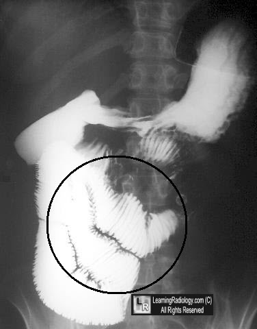
  • In progressive systemic sclerosis barium examination shows dilatation of the duodenum and jejunum, diminished peristalsis, decreased motility and delayed transit. Sacculations, also known as pseudodiverticula, are seen frequently as large, broad-based outpouchings with a somewhat squared contour.
  •  A characteristic sign of progressive systemic sclerosis is an increased number of mucosal folds – the ‘wire spring or ‘hidebound’ appearance 


Pseudomembranous colitis


eurorad.org

  • Pseudomembranous colitis results from the effects of cytoplasmic endotoxins produced by overgrowth of Clostridium difficile, usually as a result of broad-spectrum antibiotic therapy.
  • Plain radiographs may show a generalized ileus and nodular haustral thickening. 
  • CT may show gross wall thickening, with marked mucosal enhancement and extensive low attenuation from submucosal oedema. This produces a very prominent target sign, often described as the accordion sign .
  • The accordion sign  is typical of pseudomembranous colitis, but may be seen in acquired immune-deficiency syndrome (AIDS)-related or ischaemic colitis and in severe oedema from cirrhosis.