- Recent advances in MR imaging point the way to a reliable marker for Parkinson disease, which is usually diagnosed almost solely through medical history and clinical findings like muscle stiffness and tremors.
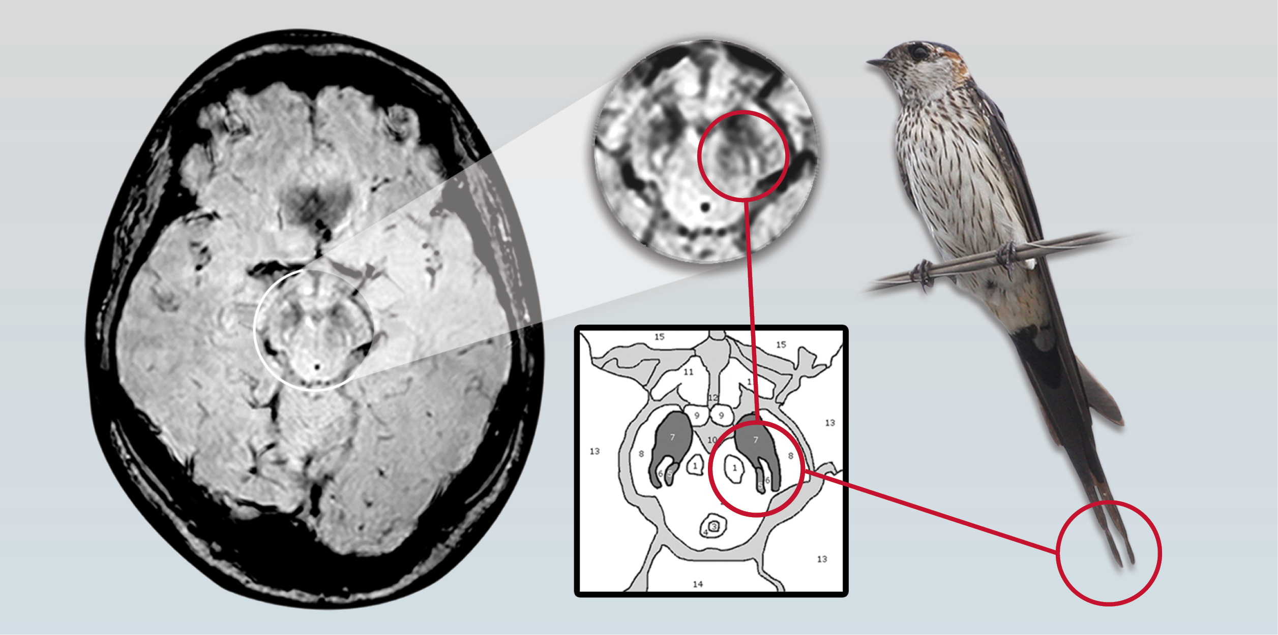
- Three MR imaging-based studies have discovered distinctive changes in the substantia nigra (SN), a crescent-shaped mass of cells in the midbrain that normally produces the neurotransmitter dopamine.
- Parkinson disease patients lose dopamine-producing cells in the SN, leading to problems with motor control among other symptoms. All three studies showed consistent differences in the appearance of the SN in normal patients compared with patients diagnosed with Parkinson disease.
- The initial study was conducted by neuroradiologists, neurologists and physicists of the University of Nottingham in the U.K., and published in the July 2013 edition of Neurology. Led by resesarchers Anna Blazejewska, Ph.D., and Stefan Schwarz, M.D., the team investigated 7-T MR imaging changes to nigrosome-1 of the substantia nigra revealing an oval shaped structure found in healthy patients but absent in patients with Parkinson disease.
- In a follow-up study by the same research team led by Dr. Schwarz, previous findings were translated to a 3-T MR imaging platform revealing a specific “swallow tail” shape indicating the presence of nigrosome-1—part of the SN that has a dense concentration of dopamine-producing cells. Researchers determined that the split-tail shape was clearly visible in normal patients but not present in the patients with Parkinson disease.
RSNA NEWS
No comments:
Post a Comment