--Fetal echo detects majority of structural cardiac abnormalities.
---most neonates born with CHD have no preidentified risk factors.
---prenatal detection rates of isolated CHD have remained below 50% in the general population
---Four chamber view and the outflow tracts are used for prenatal detection of CHD
---Outflow tracts view of fetal heart show better detection of CHD when compared to the four chamber view
---Nuchal translucency ( 10 -14 weeks) greater than or equal to 3,55 mm is an indication for fetal echo
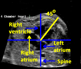
Criteria of normal four chamber view of heart
1.normal fetal situs
2.normal size in relation to chest
3.the two atria a---equal in size and the flap of the foramen ovale is seen in the left atrium
4.the venticles are equal in size and contactility ,moderator band in the apex of right ventricle
5.atrial and ventricular septae are normal appearing
6.the AV valves are normal appearing .the tricuspid valve appears to insert more apically on the ventricular septum
CARDIAC ABNORMALITIES associated with abnormal four-chamber view of the heart
1.mitral/aortic atresia
2.tricuspid /pulmonary atresia
3.Ebstein anomaly /tricuspid valve dysplasia
4.AV septal defect
5.large VSD
6.single ventricle (double inlet)
7/severe/aortic /pulmonary stenosis
8.severe coarctation of aorta
9.TAPVC
10.cardiomyopathies /tumour
CARDIAC ABNORMALITIES associated with NORMAL four-chamber view of the heart
1.TOF
2.TGA
3.double outlet right ventricle
4.small VSD
5.common arterial trunk
6.aortic arch abnormalities
No comments:
Post a Comment